NIH Awards $2.8 Million Grant to NC State Developmental Biologist for Work Investigating Organ Formation
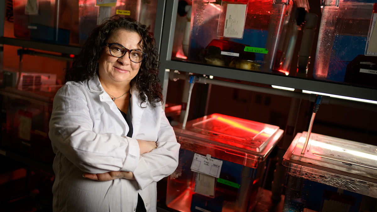
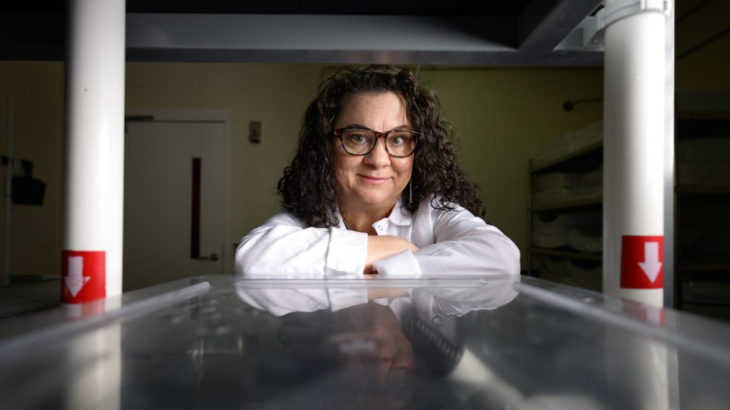
Dr. Nanette Nascone-Yoder, a professor in the NC State College of Veterinary Medicine’s Department of Molecular Biomedical Sciences, has been awarded a $2.8 million grant from the National Institutes of Health for her research investigating how cellular mechanisms guide left-right asymmetric organ formation.
Abnormal left-right asymmetry underlies some of our most common and severe birth defects. Her focus is on understanding how internal organs get their asymmetrical shapes and positions — stomach, heart and spleen on the left, liver on the right, etc. — and why those processes sometimes fail.
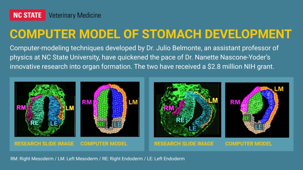
Dr. Julio Belmonte, an assistant professor of physics at NC State University, is her co-investigator on the project, using his expertise in computational modeling to provide Nascone-Yoder with a “virtual laboratory” that cuts out numerous steps and copious amounts of time from traditional research methods.
“He takes our images of organ development and makes a virtual 2D or 3D organ on the computer, complete with the appropriate numbers of cells and different layers of tissues and the appropriate geometry,” Nascone-Yoder says. “He can then program the cells to do what we think they’re doing in the embryo to see if our hypothetical mechanisms can actually produce the expected organ shape.”
Both professors are part of NC State University’s Quantitative and Computational Developmental Biology Cluster, one of 10 groups of interdisciplinary researchers hired as part of the Chancellor’s Faculty Excellence Program that brings together interdisciplinary groups of scientists to address complex challenges together.
With five years of NIH funding, the project will combine Nascone-Yoder’s experiments using frog embryos and Belmonte’s computational modeling to explore how various genetic manipulations affect organ development. The research could lead to strategies for early detection or prevention of developmental abnormalities, such as congenital heart defects, in humans and animals.
Nascone-Yoder hopes to provide crucial insights into left-right asymmetry by looking at how the stomach forms its proper curvature, a process she likens to taking a ziti and trying to turn it into a macaroni.
“We decided to start with the stomach because it’s basically a straight tube that curves in a specific direction,” she says. “When we understand the basic cellular processes that shape the development of this simple asymmetry, those processes may reveal novel insights into the development of other left-right asymmetric organs.”
The heart, for example, also begins its development as a straight tube that forms a simple curvature before acquiring additional complexity.
Nascone-Yoder previously found that each side of the embryonic stomach develops independently, yet the organ somehow integrates this information to form the appropriate overall 3D shape. Understanding how that happens is what the grant will help fund, Nascone-Yoder says.
“We understand the process that occurs prior to the organs developing that makes one side of the embryo know that it’s ‘left’ and the other side know that it’s ‘right’,” she says. “But how this information then instructs the stomach or heart to curve, and in the proper direction, we don’t really understand.”
For her in vivo experiments, Nascone-Yoder uses Xenopus laevis frog embryos, which remain transparent as their organs begin to develop. Because the very first cell division separates the embryo into left and right sides, the scientists can independently manipulate the genes on each side of the embryo to see how those changes affect subsequent organ curvature.
“We can take genes that are expressed on the left side and express them on the right side and vice versa,” she says. “We can also knock out genes on the left or right side and see if the stomach still forms the normal shape. The frog is a powerful model because it forms organs with the same laterality as mammals, including humans.”
The stomach is composed of multiple layers of tissue, and Nascone-Yoder hopes to pinpoint what each layer does. Belmonte’s computer modeling allows the scientists to mimic changes in the virtual stomach and to evaluate how they affect curvature, which validates or refutes specific hypotheses about the cellular events that occur in each tissue layer. This process continuously leads to new insights and more information to better design the next Xenopus experiment.
Belmonte will also offer his own theories about how the physical and mechanical properties of the stomach contribute to its asymmetrical development. For instance, Nascone-Yoder says, he might say he thinks the tissue layers are adhering more on one side versus the other so the stomach can curve in a particular direction, and then he would program the difference and watch how the virtual organ develops.
“And then we can go back to the frog embryo and ask, ‘Are cell adhesion genes expressed at higher levels on that side?’ If so, this would indicate that Dr. Belmonte’s theory may be correct, giving us a new hypothesis to test in vivo,” she says. “It’s pretty exciting to be able to take both an in vivo and a computational model and have them inform each other. It accelerates understanding.”
Nascone-Yoder, who obtained her Ph.D. in cell and developmental biology at Harvard University, established her laboratory at NC State’s College of Veterinary Medicine in 2006. She has received multiple grants over the years, including from the National Science Foundation, the American Heart Association and previously from the National Institutes of Health.
Dr. Joshua Stern, associate dean of research at the college, says Dr. Nascone-Yoder has pioneered some incredible developmental biology research at its most basic levels.
“She has led to our key understandings of organ development, and her program is very highly recognized,” Stern says. “For this grant, she basically put forth a conundrum to NIH, and NIH said, ‘You’re right. We need to understand that, and we think you’re the right person to help us do it.’”
This post was originally published in Veterinary Medicine News.